Table of Contents
Procedure Summary
- The surgical procedure involves six main steps: ovarian mobilisation, drainage of the ovarian endometrioma, vasopressin injection, developing the plane between the endometrioma and ovarian tissue, achieving haemostasis by suturing the base of the ovary, and temporary ovarian suspension.
- Intracorporeal knot tying is used to reapproximate the ovary.
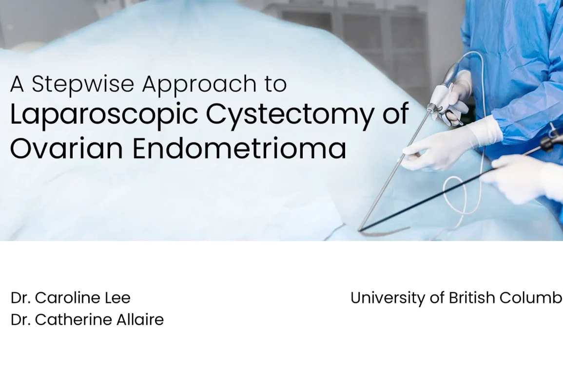
- The procedure is illustrated through a video demonstration of a case study involving a 37-year-old woman with pelvic pain due to bilateral endometriomas.
- The surgical steps are designed to minimise damage to the ovary and preserve ovarian reserve.
What is Laparoscopic Cystectomy
- Laparoscopic Cystectomy is a minimally invasive surgical procedure used to remove cysts from the ovaries.
- It involves making small incisions in the abdomen to insert a laparoscope (a thin, lighted tube with a camera) and surgical instruments.
- The cyst is carefully dissected and excised, preserving as much healthy ovarian tissue as possible.
- The procedure allows for better visualization and magnification, enabling precise removal of the cyst while minimizing damage to surrounding structures.
- Laparoscopic Cystectomy offers advantages such as reduced post-operative pain, shorter hospital stay, faster recovery, and smaller scars compared to traditional open surgery.
Presented By
Affiliations
University of British Columbia
See Also
Watch on YouTube
Click here to watch this video on YouTube.
Video Transcript
In the following video, we demonstrate a step-wise approach to laparoscopic cystectomy of ovarian endometriomas. The authors have no relevant disclosures. Here, we present a case of a 37-year-old G1P0 woman with a longstanding history of dysmenorrhea and pelvic pain in the context of known bilateral endometriomas. She was experiencing persistent pelvic pain despite current suppressive therapy with dienogest. For this reason, she is booked for conservative surgery for pelvic pain.
Surgical Steps
We’ll be illustrating a step-by-step approach to excision of ovarian endometriomas. The steps will include, one, complete mobilisation of the ovary from the pelvic side wall. Two, drainage of the ovarian endometrioma. Three, injection of dilute vasopressin into the base of the ovarian endometrioma. Four, completion of the cystectomy by developing the plane between the endometrioma and ovarian tissue, using blunt dissection and limited use of energy. And five, achieving haemostasis by suturing the base of the ovary in a purse string fashion.
As a final consideration, a temporary ovarian suspension should be completed in the event of extensive underlying peritoneal dissection. This step will help prevent post-operative adhesion formation.
Ovarian Mobilisation
Upon laparoscopic entry, we made note of a large right-sided endometrioma. With this dissection, you’re going to develop that plane between the ovary and the pelvic side wall. Rod may be inserted parallel to this plane. Allow for efficient blunt dissection. Such movement can be done circumferentially until the ovary is completely free. During the process of ovarian mobilisation, the ovarian endometrioma is often entered. The ovarian endometrioma is then completely drained.
Vasopressin Injection
For step three, we prepared a solution of 20 units of vasopressin in 50cc of normal saline. This solution was injected into the base of the endometrioma to allow for hydrodissection. With the aid of the vasopressin, we were able to find an avascular plane between the endometrioma and ovarian tissue. As demonstrated in our video, we used a combination of traction, countertraction, and blunt dissection in order to develop our plane. We set it on the side of easiest access, and circumferentially released the endometrioma from its space until it was completely free.
In our next endometrioma, the plane was not as clearly demarcated. Due to our concerns about haemostasis, we made the decision to use our harmonic device. This device allowed us to deliver focused energy to develop our planes. This approach allowed us to maintain as much normal ovarian tissue as possible while optimising haemostasis. We noted the presence of a final endometrioma. This was removed using the same techniques described previously.
Suturing the Base of the Ovary
With step four completed, we needed to achieve haemostasis by suturing the base of the ovary. Due to our exclusive use of 5mm trocars, we used a backloading technique to introduce the suture under direct visualisation. This approach allowed us to safely introduce the needle while avoiding the use of larger trocars.
The needle was then loaded in the usual fashion, and a purse string approach to suturing was used to achieve haemostasis. We adapted the suturing technique to minimise the impact of surgery on ovarian reserve. The rationale for this technique was provided by Wang and colleagues, as published in February 2019 in the article titled, Effect of laparoscopic endometrioma cystectomy on AMH levels. The suturing technique was found to be associated with a lower post-operative decrease in AMH level compared to coagulation surgery.
After completion of our purse string suture, we noted that the ovary was reapproximating appropriately. Using irrigation, we completed a final check for haemostasis. Intracorporeal knot tying was used to reapproximate the ovary. As haemostasis had been achieved, we were able to cut our laparoscopic suture. It was then removed under direct visualisation.
The final step is demonstrated here in a separate video. We begin by introducing the Keith needle into the peritoneal cavity under direct visualisation. The needle is then positioned underneath the ovary and used to pierce the ovarian cortex. The needle is then removed from the peritoneal cavity via the anterior abdominal wall. The suture is secured externally and removed between post-op day number five and seven.
Conclusion
At the conclusion of our surgery, our operative findings were stage IV endometriosis with bilateral endometriomas and superficial endometriosis of the posterior cul-de-sac. The procedure performed included excision of bilateral endometriomas and excision of cul-de-sac peritoneal endometriosis. These additional steps were not included in the video.
In summary, the steps of an ovarian endometrioma excision include, one, complete mobilisation of the ovary. Two, drainage of the ovarian endometrioma. Three, injection of vasopressin into the base of the endometrioma. Four, developing the plane between the endometrioma and ovarian tissue. Five, achieving haemostasis by suturing the base of the ovary. And six, considering a temporary ovarian suspension of the affected ovary.


