Table of Contents
- Procedure Summary
- Authors
- Youtube Video
- What is a Trachelectomy?
- What are the risks of Trachelectomy?
- Video Transcript
Procedure Summary
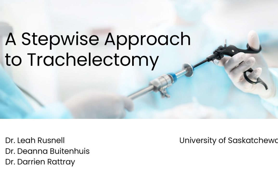
- The video presents a laparoscopic approach to trachelectomy.
- Trachelectomy is sometimes performed for patients who have previously undergone a hysterectomy.
- Trachelectomy is performed in five basic steps: restoring anatomy, creating a bladder flap, performing anterior and posterior colpotomy, lateralizing the uterine artery pedicles, and closing the vaginal cuff.
- The patient in the video underwent a supracervical hysterectomy before deciding on trachelectomy.
- The patient’s dyspareunia improved following surgery.
Presented By


Affiliations
See Also
Watch on Youtube
Click here to watch this video on YouTube.
What is a Trachelectomy?
- Trachelectomy is a surgical procedure that involves the removal of the cervix while preserving the uterus
- It is typically performed as an alternative to a radical hysterectomy, allowing women to maintain their fertility and the possibility of future pregnancy.
- The procedure involves removing the cervix and some surrounding tissue, while leaving the upper part of the uterus intact.
- The main goal of trachelectomy is to treat cervical cancer while preserving reproductive options for women of childbearing age.
What are the risks of Trachelectomy?
Risks may include:
-
Bleeding or Hemorrhage: Excessive blood loss during or after the surgery.
-
Infection: Risk of postoperative infection in the surgical area or pelvic cavity.
-
Damage to Surrounding Organs: Possibility of injury to bladder, ureters, or intestines.
-
Cervical Stenosis: Narrowing of the remaining cervical canal, potentially affecting fertility or menstrual flow.
-
Sexual Dysfunction: Potential for pain during intercourse or changes in sexual function.
-
Preterm Labor: Increased risk of preterm labor in future pregnancies due to a weakened cervical structure.
-
Miscarriage or Pregnancy Complications: Elevated risk due to the changed structure and function of the cervix.
-
Anesthesia Risks: Side effects or complications related to the anesthesia used during the procedure.
-
Emotional Impact: Psychological stress or anxiety related to cancer treatment and fertility concerns.
-
Recurrence of Cancer: While the goal is to remove all cancerous tissue, there’s a risk that some cells may remain, leading to recurrence.
Consultation with a qualified healthcare provider is essential for a full understanding of the risks and whether a trachelectomy is the most appropriate treatment option for your condition.
Video Transcript
In the following video we present a laparoscopic approach to trachelectomy. The authors have no relevant disclosures. A supracervical approach is undertaken in about 10% of all hysterectomies. A supracervical approach may be favoured in a variety of situations. Some examples are to lower the risk of bladder injury in the setting of severe bladder adhesions at the time of unplanned hysterectomy for a postpartum haemorrhage or patient preference.
However, about 25% of patients who undergo supracervical hysterectomy will continue to have bothersome symptoms. Many of these women will go on to have a trachelectomy. Studies have not demonstrated a difference in complication rate between supracervical or total hysterectomy. Overall, the benefits of supracervical hysterectomy remain largely theoretical.
However, some women will still choose preservation of the cervix. Many of these women hold the belief that sexual function and satisfaction will be negatively affected if the cervix is removed. However, this is not demonstrated in the literature. Patients should be counselled that the best predictor of postoperative sexual function is a patient’s preoperative sexual function.
Patients with Previous Hysterectomy
Trachelectomy for patients who have previously undergone a hysterectomy involves the five following steps. Step one, restore anatomy. Depending on medical and surgical history, there may be adhesions of small or large bowel, rectum, bladder or adnexa to the cervix. Adhesiolysis and restoration of anatomy must be performed initially for visualisation of the cervix.
Step two, create a bladder flap. Step three, perform anterior and posterior colpotomy. Step four, lateralise the uterine artery pedicles. The colpotomy can then be completed. Step five, close the vaginal cuff. In this video we present the case of a 50-year-old G4T2 woman who is otherwise healthy. She underwent hysterectomy for issues of heavy menstrual bleeding and dyspareunia. She preferred a supracervical approach for personal reasons. Unfortunately, her dyspareunia persisted and five years later she decided to proceed with trachelectomy.
Step 1
Step one, restore anatomy. A survey of a pelvis is performed. Fortunately, despite our patient’s previous surgeries, there are no adhesions of any structures to the cervix in this case. The ureters are visualised transperitonealy and are far from our site for colpotomy. Ureterolysis is not necessary in this case. A sponge-stick is placed in the vagina to delineate the cervix.
Step 2
Step two, create a bladder flap. The vesicovaginal space is developed and the bladder should be deflected inferiorly, about two centimetres from the site of planned colpotomy. Here, once we know the bladder is not overlying the cervix, the internal os is perforated with a dilator. A uterine manipulator with a cervical cup is then placed.
By using the intrauterine tip, we ensure a snug fit of the cervical cup, so that cervix will be removed entirely, while maintaining vaginal length. Here we can see the cervix well delineated and the inferior margin of the bladder that needs to be further mobilised to facilitate the anterior colpotomy. Both blunt and sharp dissection are used to further deflect the bladder off of this pubocervical fascia, until the site for colpotomy is clear.
Step 3
Step three, anterior and posterior colpotomy. The monopolar hook is used to perform the anterior and posterior colpotomy. By performing this step first, the location of the uterine vessels is well delineated.
Step 4
Step four, lateralise the uterine artery pedicles. Since the anterior and posterior colpotomies have already been performed, we now only need to lateralise the uterine artery pedicles as far as necessary to complete the colpotomy. The bipolar vessel ceiling device is used to desiccate and lateralise the uterine artery pedicle and the colpotomy is then completed. This is performed bilaterally. The cervix is removed vaginally. Section irrigation is performed and bleeding of the vaginal cuff is addressed. We try to use as little energy as possible.
Step 5
Step five, close the vaginal cuff. We close the vaginal cuff with a barbed suture in a running unlocked fashion for good anatomic and haemostatic effect.
Surgical Outcome
The patient was discharged home the same day and her recovery was unremarkable. Her dyspareunia improved following surgery. Pathology confirmed benign cervical tissue. Trachelectomy is sometimes considered an intimidating procedure. Our case helps to demonstrate that trachelectomy is potentially within the scope of any practitioner performing total laparoscopic hysterectomies. Although this case demonstrates a simple and straightforward trachelectomy, these same steps can be applied to more complex cases as well.
Conclusion
In summary, trachelectomy following a previous hysterectomy is performed with five basic steps. One, restore anatomy. Two, create a bladder flap. Three, anterior and posterior colpotomy. Four, lateralise the uterine artery pedicles. The colpotomy can then be completed. Five, close the vaginal cuff.


