Table of Contents
Video Description
The objectives of this video are to define bowel endometriosis and to explore various surgical parameters for the different types of surgical excision.
Then, a specific surgical approach will be demonstrated. When planning a surgical approach to deep endometriosis of the bowel, patient characteristics such as age and BMI, as well as their specific symptoms and level of pain, quality of life and fertility goals must be considered.
As well, the actual lesion must be investigated with respect to size, number, location, depth of infiltration, and amount of intestinal wall circumference involved. Then, various surgical techniques can be performed depending on these specific characteristics, such as nodule shaving, nodular resection and segmental resection and re-anastomosis.
A surgical case is then utilized to demonstrate a nerve sparing and blood supply conserving technique of segmental resection after intra-operative sigmoidoscopy demonstrated luminal obstruction.
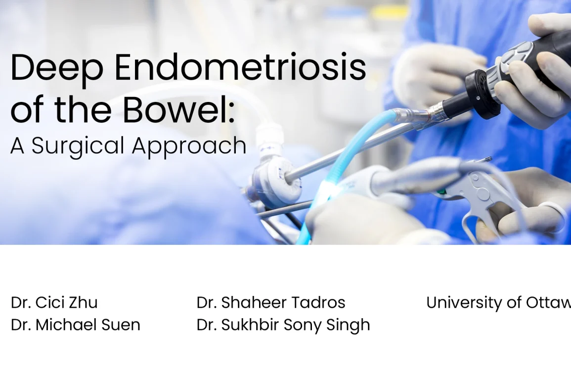
Presented By
Affiliations
University of Ottawa
See Also
Watch on YouTube
Click here to watch this video on YouTube.
What is Deep Endometriosis of the Bowel: A Surgical Approach?
Deep Endometriosis of the Bowel: A Surgical Approach refers to the surgical strategies used to treat a severe form of endometriosis that affects the bowel.
-
Deep Endometriosis:
- A severe form of endometriosis where endometrial-like tissue grows beneath the peritoneum, often affecting the bowel and other pelvic organs.
- It can lead to significant pain, infertility, and bowel-related symptoms like irregular bowel movements, bloating, and rectal bleeding.
-
Surgical Approach:
- Surgery is considered when medication and other non-invasive treatments fail to relieve symptoms.
- The goal is to remove endometrial implants while preserving the bowel and surrounding structures.
What are the Risks of Deep Endometriosis of the Bowel: A Surgical Approach?
The surgical approach to treating deep endometriosis of the bowel, while often necessary, carries several risks. Risks may include:
-
Bowel Perforation: The risk of accidentally creating a hole in the bowel wall during surgery, leading to potential infection or sepsis.
-
Bleeding: Heavy bleeding can occur during or after surgery, possibly requiring a blood transfusion.
-
Infection: As with any surgery, there’s a risk of infection at the incision site or within the pelvic cavity.
-
Organ Damage: Near by organs, such as the bladder, ureters, or other sections of the bowel, may be inadvertently damaged during surgery.
-
Adhesions: Scarring can occur post-surgery, leading to tissue and organ adhesion, which might cause pain or bowel obstruction.
-
Anastomotic Leakage: If bowel resection and reconnection (anastomosis) are performed, there’s a risk of leakage from the site where the bowel segments are joined.
-
Changes in Bowel Function: Temporary or permanent changes in bowel habits, including constipation or diarrhea, may occur post-surgery.
-
Fertility Issues: While surgery can improve fertility in some individuals with endometriosis, potential damage to the reproductive organs can occur, affecting fertility.
-
Recurrence: Endometriosis may recur even after surgery, necessitating further treatment or additional surgical procedures.
Each patient’s risk profile may differ based on their overall health, the extent of the disease, and the specific surgical technique employed. Thorough pre-surgical assessment and planning, as well as post-operative care, are crucial in mitigating these risks.
Video Transcript: Deep Endometriosis of the Bowel: A Surgical Approach
Deep endometriosis of the bowel, a surgical approach by the Minimally Invasive Gynecology group at The Ottawa Hospital. The objectives of this video are to define bowel endometriosis and explore various surgical parameters for the different types of surgical excision. Then a specific surgical approach will be demonstrated.
The bowel is a common site of nonreproductive organ endometriosis with 90% of lesions involving the colon. With an incidence up to 12% in women with endometriosis, it most commonly affects the bowel serosa, but can invade deeper into the muscularis propria and even the lumen.
Transvaginal ultrasound is the most sensitive modality for preoperative detection of endometriosis involving bowel. If symptomatic, most reported symptoms include dyschezia, rectal bleeding, and obstructive symptoms such as constipation, faecal urgency and tenesmus.
Surgical management of bowel endometriosis is indicated for symptom relief, intolerance to medical management, and to prevent complete obstruction. For deep endometriosis it often involves a multidisciplinary team of minimally invasive gynaecologists and colorectal or general surgeons.
When planning a surgical approach to deep endometriosis of the bowel, patient characteristics as well as their specific symptoms and level of pain, quality of life, and fertility goals must be considered. As well, the actual lesion must be investigated with respect to size, number, location, depth of infiltration, and amount of intestinal wall circumference involved.
Then various surgical techniques can be performed depending on these specific characteristics, such as nodule shaving, nodular resection, and segmental resection and reanastomosis.
Importantly, surgeons must acknowledge the potential surgical complexity of these cases and counsel towards both surgical and nerve related complications.
The following case demonstrates an approach to segmental resection. Our surgical case is of a 34-year-old nulliparous woman with a known history of deep endometriosis who had presented for surgical management. She also had a previously undisclosed two-year history of increasing rectal bleeding and more recent faecal urgency.
Initial laparoscopic exploration revealed endometriosis throughout the pelvis including the anterior bladder flap, both pelvic side walls, and uterosacral ligaments.
Once this was all resected, examination of the bowel did reveal a significant 4 to 5 cm nodule of disease at the rectosigmoid junction.
First, overlying fat and mesentery was carefully dissected away from the lesion and a suture was placed at the nodule as delineation for a potential full thickness disc excision.
Sigmoidoscopy was then performed intraoperatively in order to delineate any luminal disease. During its implementation, it became obvious that the lesion was causing a stricture with luminal narrowing and that the scope was unable to pass beyond the endometriotic nodule.
The stricture was too significant for shaving or nodular resection. Therefore, a decision was made to complete segmental resection and reanastomosis for full symptomatic relief.
Starting on the patient’s left, peritoneum was opened high on the mesentery given the benign nature of the disease. Retroperitoneal dissection clearly reveals the left ureter which was carefully delineated and lateralised.
The inferior mesenteric artery provides blood supply to the sigmoid colon as well as the rectum via its terminal branch, the superior rectal artery. Closed dissection of terminal vessels prevents damage to and preserves larger structures.
Sympathetic enervation to the rectosigmoid junction arises from the inferior hypogastric plexuses providing overall inhibitory response. Parasympathetic enervation arises from the pelvic splanchnic nerves from S2, 3, 4 providing a stimulatory response.
By avoiding the tissue where all these nerves lie in our dissection, we preserve full function and avoid complications including faecal incontinence and voiding dysfunction.
Once dissection is complete, distal bowel transection was then completed with one firing of a 60 mm Endo GIA stapler.
The sigmoid colon was then mobilised to the splenic flexure to gain length and mobility by transecting medial peritoneal attachments and distal sigmoid mesentery. The inferior mesenteric artery was preserved.
A 4 cm mini-laparotomy was then made to extract the specimen and a 31 mm anvil was secured into the healthy proximal bowel with a 2-0 prolene purse string suture, and then the colon was replaced.
Pneumoperitoneum was reestablished and anastomosis was created without tension with the EEA 31 mm just above the horizontal staple line of the rectum. Anastomosis appeared tension free and well vascularised.
At her postoperative follow up, the patient was able to report resolution of rectal symptoms.
In summary, we described preoperative surgical parameters that help guide surgical management of bowel endometriosis and used a surgical approach to highlight relevant anatomy. Thank you to our great team of collaborators.