Laparoscopic Cervical Myomectomy with Pre-operative Uterine Artery Embolization and Concomitant Cerclage

Last updated:

Table of Contents

Video Description

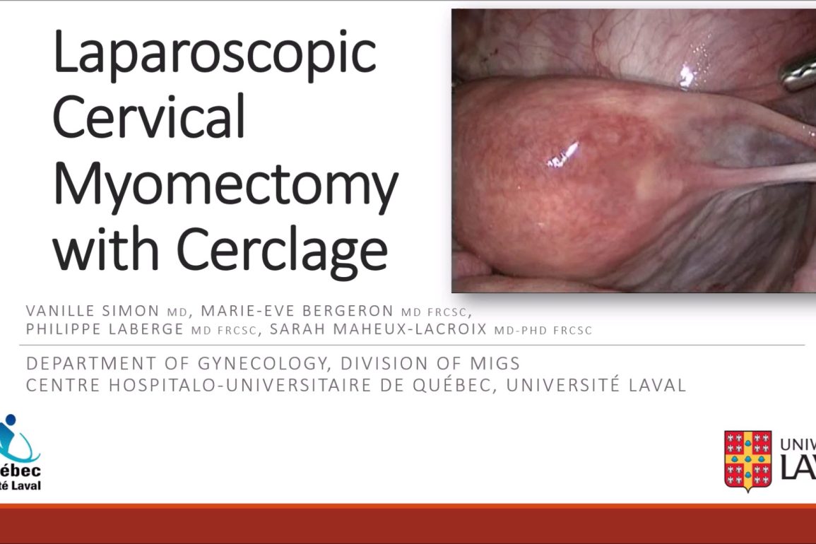
Cervical myomectomy is a surgical challenge and the risk of subsequent cervical incompetence is unknown. We presented the case of a 30-year-old woman, nulligravida, with a 12 cm cervical leiomyoma, who consulted for heavy menstrual bleeding and pelvic pain.

After failure of multiple medical therapies, a laparoscopic cervical myomectomy was successfully performed. Adjuvant pre-operative uterine artery embolization with gelatin sponges was used to reduce surgical blood loss, as an alternative to intra-operative ligation of uterine arteries when access to the retroperitoneum is limited by the size and location of leiomyomas.

In order to prevent cervical incompetence, a concomitant laparoscopic cerclage was achieved since the integrity of the cervix has been compromised by the myomectomy.

Presented By

Female doctor avatar
Dr. Vanille Simon
Female doctor avatar
Dr. Marie-Eve Bergeron
male doctor avatar
Dr. Philippe Laberge
Female doctor avatar
Dr. Sarah Maheux-Lacroix

Affiliations

Université Laval, Centre Hospitalier Universitaire de Québec

Watch on YouTube

Click here to watch this video on YouTube.

What is Uterine Artery Embolization and Concomitant Cerclage?

Laparoscopic cervical myomectomy with pre-operative uterine artery embolization (UAE) and concomitant cerclage is a specialized procedure for treating cervical fibroids, particularly in women seeking to preserve fertility. UAE is first performed to decrease blood supply to the fibroids, reducing their size and minimizing blood loss during surgery. Following UAE, laparoscopic myomectomy is carried out using a camera and instruments inserted through small abdominal incisions to remove the fibroids. The procedure is minimally invasive, aimed at reducing recovery time and surgical risks. Additionally, a cerclage is placed around the cervix to prevent premature opening, supporting pregnancy maintenance post-surgery. This comprehensive approach not only addresses the removal of fibroids but also enhances the structural integrity of the cervix, crucial for future pregnancies. The combination of these techniques offers an effective solution to manage cervical fibroids while optimizing fertility outcomes.

What are the Risks of Uterine Artery Embolization and Concomitant Cerclage?

The combination of laparoscopic cervical myomectomy with pre-operative uterine artery embolization (UAE) and concomitant cerclage carries specific risks, alongside the general benefits of treating cervical fibroids and supporting fertility. These risks may include:

  • Infection: Surgical procedures and uterine manipulations increase the risk of pelvic infections.
  • Bleeding: Despite the reduced blood flow from UAE, significant bleeding can occur during myomectomy.
  • Injury to Surrounding Organs: Laparoscopy involves the risk of accidental damage to nearby organs such as the bladder or intestines.
  • Uterine Scarring: Any uterine surgery can lead to scarring, which may affect fertility and future pregnancies.
  • Cerclage Complications: The cerclage can cause cervical irritation, premature labor, or infection.
  • Recurrence of Fibroids: There’s a possibility of fibroid recurrence, requiring further treatment.
  • Failure of Procedure: In some cases, the procedures may not fully resolve symptoms or could negatively impact fertility.
  • Risks from Embolization: UAE can lead to complications such as post-embolization syndrome, characterized by pain, fever, and nausea.

Patients considering this treatment should discuss these risks in detail with their healthcare provider to make an informed decision based on their specific health situation and reproductive goals. The multidisciplinary approach aims to maximize benefits while minimizing complications, especially in women desiring to maintain or improve fertility.

Video Transcript: Uterine Artery Embolization and Concomitant Cerclage

This video presents a laparoscopic cervical myomectomy with cerclage. The authors declare no disclosure. This is the case of a 30-year-old woman who consulted for severe, heavy menstrual bleeding caused by a large cervical myoma of 12 centimetres with a right ureterohydronephrosis. The patient was in good health and had never been sexually active or pregnant. The leiomyoma was filling her pelvis and was palpable at only a few centimetres of the vaginal introitus, with a cervical os difficult to identify.

She was referred to our mix department after failure of multiple medical therapies, including tranexamic acid, ulipristal acetate, leuprolide acetate, and failure of interventional therapy with UAE. A decision was made to perform laparoscopic cervical myomectomy with concomitant cerclage of the cervix. We will review three main considerations for cervical myomectomies, which are, one, prevention of bleeding. Two, prevention of injury to surrounding structures, specifically the ureters. Three, evaluation of the need for cervical cerclage.

The risk of bleeding is significant during cervical myomectomy despite occlusion of uterine arteries, as cervical leiomyoma are often vascularised by branches of the vaginal arteries. In this context, several actions should be combined to reduce the risk of bleeding, including intravenous tranexamic acid, intrarectal misoprostol, sub-serosal vasopressin and occlusion of uterine arteries. In this case, intraoperative occlusion of uterine arteries with clips or with penrose drain was expected to be difficult given the size and localisation of the leiomyoma.

 Therefore, a decision was made to use preoperative uterine artery embolization with gel form. The day of the surgery, all members of the surgical team were aware of the potential risk of significant bleeding and conversion to laparotomy, and blood products were available. Cervical leiomyomas are very close to important structures and anatomy is often disturbed by their presence. It is therefore mandatory to make sure to, one, dissect the bladder down. Two, identify the ureters and bowel.

Three, identify the capsule of the leiomyoma and the correct plan of dissection. Myomectomy is achieved by applying traction with a tenaculum and by applying countertraction using an atraumatic grasper. At the end of the myomectomy, it became clear that the cervical integrity was compromised. Here, as you can see, with the anterior vaginal wall open and the cervical OS on the right, only a few stretch fibres of cervix remain on the left.

Knowing that there is an important risk of cervical incompetence following cervical surgeries and that prophylactic abdominal cerclage is indicated during trachelectomies, a decision was made to perform a concomitant cerclage with the patient’s preoperative consent. First, the bladder had already been dissected down and the broad ligament is fenestrated on both sides. Second, uterine arteries are well identified and skeletonised. This is an essential step, as we will pass the needle medially to the uterine vessels.

Third, using a prolene zero, two loops of suture are placed around the cervix at the height of the cervicoisthmic junction. We have to be cautious not to occlude the uterine arteries. The level of the cerclage is determined by identifying the utero-sacral ligaments posteriorly and the vaginal fornix anteriorly. The knot is tied with enough tension, just enough to create a visible depression in the soft tissue, but without generating a strangulation or an ischaemia. The vagina is closed with a running suture using V-loc zero.

Then the fibres of the cervix are reapproximated with a continuous locking stitch using a V-loc zero. Finally, the anterior peritoneum is closed with a purse-string suture using a zero Monocryl to prevent the incarceration of other organs and to avoid future development of adhesions. At the end of surgery, important structures have been preserved and the cerclage is well positioned with adequate tension. The patient was informed that she will require caesarean section due to the cerclage, but also given the risk of uterine rupture after this myomectomy.

In conclusion, for cervical myomectomy. One, adjuvant pre-operative UAE with gel form can be considered, especially when access to uterine arteries is expected to be difficult given the size and localisation of the leiomyoma. Two, ureters and uterine arteries need to be carefully identified because of an anatomic distortion making them even closer. Three, consider performing an abdominal cerclage when the integrity of the cervix has been compromised by the myomectomy.