Table of Contents
- Procedure Summary
- Authors
- Youtube Video
- What is Managing the Adherent Bladder at the Time of Total Laparoscopic Hysterectomy?
- What are the Risks of Managing the Adherent Bladder at the Time of Total Laparoscopic Hysterectomy?
- Video Transcript
Video Description
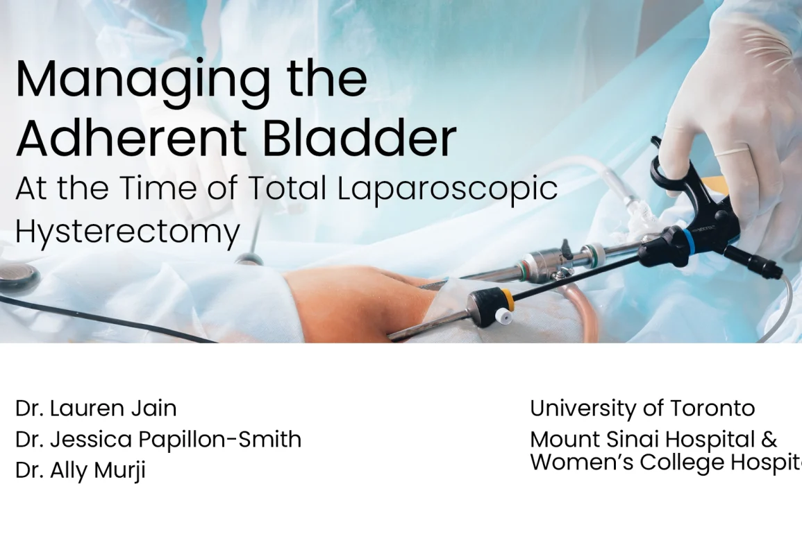
This video discusses tips and tricks in dissecting an adherent bladder safely at the time of total laparoscopic hysterectomy.
Presented By
Affiliations
University of Toronto, Mount Sinai Hospital & Women’s College Hospital
Watch on YouTube
Click here to watch this video on YouTube.
What is Managing the Adherent Bladder at the Time of Total Laparoscopic Hysterectomy?
Managing an adherent bladder during a total laparoscopic hysterectomy (TLH) involves addressing the challenges posed by bladder adhesions to the uterus, which can complicate the surgery and increase the risk of injury.
-
Adherent Bladder: This condition occurs when the bladder is attached to the uterus due to prior surgeries, inflammation, or endometriosis, making it difficult to separate the organs during surgery.
-
Causes: Previous Cesarean sections, pelvic surgeries, or conditions like endometriosis and pelvic inflammatory disease often lead to the formation of adhesions.
-
Symptoms/Challenges: The main surgical challenge is safely dissecting the bladder from the uterus without causing damage, as adhesions increase the risk of bladder injury during TLH.
-
Surgical Approach: Management involves careful preoperative imaging to evaluate the extent of adhesions, followed by precise dissection during surgery to avoid injury to the bladder and surrounding tissues.
-
Importance of Technique: Success in managing the adherent bladder relies on meticulous surgical technique and often involves the use of specialized tools to minimize the risk of complications.
What are the Risks of Managing the Adherent Bladder at the Time of Total Laparoscopic Hysterectomy?
Video Transcript: Managing the Adherent Bladder at the Time of Total Laparoscopic Hysterectomy
This video reviews the risk factors for difficult bladder dissection at the time of total laparoscopic hysterectomy, and demonstrates an approach to dissection aimed at minimising bladder injury. Furthermore, techniques that may assist with intraoperative identification of bladder injury are highlighted.
Urologic injury at the time of total laparoscopic hysterectomy can result in significant morbidity and medicolegal implications. Specifically, it has been estimated that ureteric injury represents 17% of non-obstetric medicolegal actions in Canada.
The rate of bladder injury at the time of total laparoscopic hysterectomy has been cited at between 1.3% and 4%, whereas rates are less than 0.5% in other modalities. The majority of these injuries are along the posterior wall, and bladder adherence to the lower uterine segment is a predisposing factor.
Risk factors include endometriosis, prior caesarean section, previous pelvic surgery, fibroids, and surgeon experience. I will now review an approach to difficult bladder dissection.
The general approach to dissection of an adherent bladder includes the identification of safe dissection plans, the delineation of the bladder edges, and careful use of various laparoscopic instruments. Finally, it is important to identify any bladder or ureteric injury intraoperatively.
This video demonstrates a challenging bladder dissection at the time of total laparoscopic hysterectomy in a patient with four previous caesarean sections. As you can see, the bladder is very adherent to the uterus and anterior abdominal wall.
Upon encountering this scenario, one would intuitively want to begin dissecting in the middle. However, given that the bladder is tethered and the margins are obscured, dissection must be done cautiously, in a systematic fashion. It is important to begin dissecting in a region with normal tissue planes and to work in a lateral to medial direction towards the abnormal scarred planes. Together, these strategies help decrease the risk of bladder injury.
We begin by opening the perivesical space, and we uncover the lateral boundaries of the bladder. This enables maximal visualisation, enhanced with a 30-degree camera, as we systematically dissect the adherent bladder.
Dissection is carried out in a lateral to medial direction, and we enter here just anterior to the round ligament. Concurrent pushing and spreading opens the avascular space nicely. Here, we identify the obliterated umbilical ligament, which forms the lateral border of our space of interest. It is bound by the bladder medially, the obliterated umbilical ligament laterally, the pubic symphysis anteriorly, and the cardinal ligament posteriorly.
As can be seen here, we have an assist pulling on the Foley to identify the bladder. This forms the medial border of our dissection. After dissecting the perivesical space on the right and identifying the lateral margin of the bladder, the same is done on the left-hand side, carrying out the dissection in a lateral to medial fashion.
As shown on the right, we open the perivesical space just anterior to the round ligament. Here we can see the location of the inferior epigastric vessels. We identified the obliterated umbilical ligament, the lateral border of our space of interest. We again demonstrate the use of both pushing and spreading to open the avascular plane.
Another technique to demarcate the bladder borders is hydrodissection, the delivery of fluid into the tissue. This clip was taken from another case involving the dissection of a challenging adherent bladder. When the round ligament is transected, we place the suction irrigator just under the vesicouterine peritoneum.
Fluid follows the path of least resistance, and here we can see the avascular space between the bladder and lower uterine segment opening. Opening the perivesical space gives us the ability to appreciate the lateral bladder margins, which can be further highlighted through the use of a technique called cystosufflation. We then detach the Foley bag and insufflate the bladder with CO2 gas. This inflates the bladder and identifies the superior-most aspect where it meets the uterus.
Another technique useful in delineating the bladder margin is through the use of a bladder probe. This segment was taken from another case with a similarly adherent bladder. Here we have a cervical dilator placed through the urethra into the bladder, and we gently manipulate the probe to demarcate the edges.
For dissection, a 30-degree scope can be very useful. This allows optimal visualisation of a challenging angle. Here, you can see the subtle angle change as the assistant manipulates the camera cord.
To review, we can see that the perivesical space has been opened and the area of interest has been exposed. The specific borders, the bladder medially, the obliterated umbilical ligament laterally, the pubic symphysis anteriorly, and cardinal ligament posteriorly are clearly demarcated.
Careful dissection of an adherent bladder can be undertaken with a combination of instruments up to the surgeon’s judgement. For our dissection, we used a combination of monopolar energy, bipolar energy with tissue sealing devices, as well as sharp and blunt dissection.
As can be seen here, we begin our approach with monopolar cautery, using the L-hook. Tissue sealing devices may be used both to identify planes with blunt dissection and to take down dense adhesions with attention to haemostasis.
Here, we can see that blunt dissection can be carried out with a curved grasper, again, always working lateral to medial. Here, we can see the cervical cup is becoming more visible, and this angle is achieved with a 30-degree scope. We then change our focus and continue with our dissection from the frontal aspect of the uterus.
As we proceed with dissection towards the bladder, we continually repeat the use of cystosufflation and/or the placement of a bladder probe to enable competent, safe dissection.
As we approach the bladder, we minimise the use of cautery and switch to sharp instrumentation. Here, we can see both blunt and sharp dissection is done with the scissors.
Here, we can see the cervical cup nicely, and the bladder is well dissected. It is important that the bladder isn’t dissected too low on the vagina, as this may compromise the ureterovesical junction. We then cystosufflate the bladder once again and confirm that our plane of dissection is free of the bladder. It is important to note that this move does not confirm a lack of injury.
After difficult dissection, a cystoscopy is recommended, as the literature demonstrates a fivefold increase in detection rate of urologic injury. Secondly, if air is detected in the Foley bag, this is certainly a clue that there has been a breach to the bladder mucosa. Finally, use of methylene blue can be useful when injury is suspected.
This segment was taken from another case where we have an obvious injury with a visible Foley catheter. Here, we demonstrate the use of methylene blue to localise a suspected defect.
In summary, key principles for dissection of an adherent bladder include the identification of safe dissection planes, the delineation of the bladder edges, as well as the careful use of various laparoscopic instruments. Intraoperative identification of bladder or ureteric injury is paramount, and cystoscopy is an important player in this process.


