Table of Contents
- Procedure Summary
- Authors
- Youtube Video
- What is Surgical Management of Interstitial Ectopic Pregnancy?
- What are the Risks of Surgical Management of Interstitial Ectopic Pregnancy?
- Video Transcript
Video Description
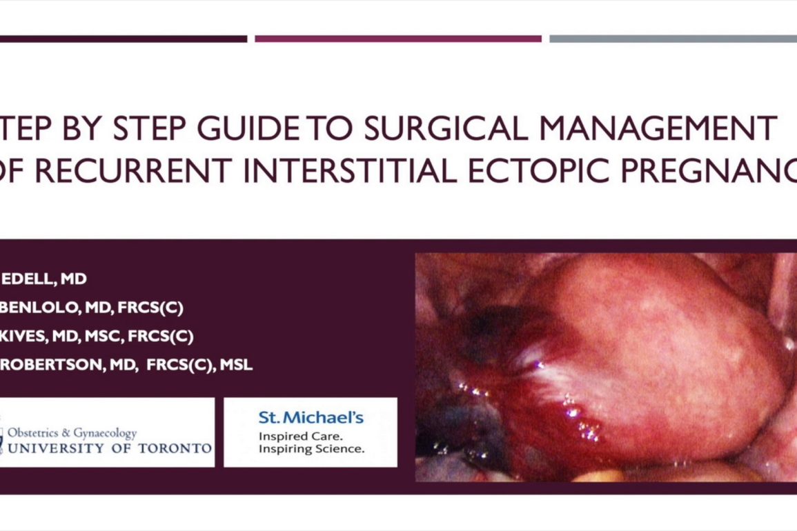
The purpose of this educational video is to provide a brief overview of interstitial ectopic pregnancy, describe a rare case of a recurrent interstitial ectopic pregnancy after previous ipsilateral cornuectomy and demonstrate a minimally invasive surgical approach to management.
We describe the case of a 38 year old G5P2 woman who presented with imaging concerning for a left interstitial ectopic pregnancy. She had previously undergone a left salpingectomy and left uterine wedge resection for separate pregnancies making the case complex and clinically fascinating.
While recurrent interstitial ectopic pregnancy does pose a high risk to patients, it can be safely managed with a minimally invasive surgical approach when techniques focused on surgical planning, blood conservation, vigilant post operative care and extensive patient counseling are implemented
Presented By
Affiliations
University of Toronto
Watch on YouTube
Click here to watch this video on YouTube.
What is Surgical Management of Interstitial Ectopic Pregnancy?
What are the Risks of Surgical Management of Interstitial Ectopic Pregnancy?
Video Transcript: Surgical Management of Interstitial Ectopic Pregnancy
This video provides a step-by-step guide to the surgical management of a recurrent interstitial ectopic pregnancy after previous cornuectomy. We will provide a brief overview of interstitial ectopic pregnancy, describe the clinical course of our patient, and outline the surgical procedure performed to manage this rare presentation.
In a normal intrauterine pregnancy, the blastocyst implants in the endometrial lining of the uterus. An ectopic pregnancy is defined as implantation at any other location. The fallopian tube is the most common site of ectopic pregnancy, accounting for 95% of cases. The other 5% can be found in the ovary, peritoneal cavity, a prior uterine scar, or the cervix.
This video focuses on interstitial ectopic pregnancy. While these are among the most rare type of ectopic, they are the most dangerous. They have a mortality rate of one in 50, seven to eight times higher than other forms of ectopic. Diagnosis is notoriously difficult. Specific radiologic criteria have been created to help aid in diagnosis.
Risk factors for interstitial ectopic pregnancies are the same as other ectopic pregnancies. Our patient is a healthy 38-year-old G5P2, who presented at eight plus one weeks gestational age, with a dating ultrasound suspicious for left ectopic pregnancy.
Her obstetrical history was significant for a spontaneous vaginal delivery, a left ectopic pregnancy managed with salpingectomy, a second spontaneous vaginal delivery, and a left interstitial ectopic pregnancy managed by cornual excision.
Dating ultrasound revealed a left extraovarian mass measuring seven plus two weeks gestational age, with a positive foetal heart rate. Her beta HCG was 87,035. The patient was completely asymptomatic, and this was a highly desired pregnancy. An MRI confirmed an interstitial ectopic pregnancy at the left uterotubal interface with an intact junctional zone.
What is most fascinating about our patient is that she had previously had a salpingectomy and cornuectomy on the same side in which she was presenting with an interstitial ectopic pregnancy. Exactly how this implantation occurred is a clinical conundrum. Medical management is reserved for patients meeting strict criteria. Approach to surgery can be conservative or more radical, with any signs of rupture being an emergency. Our patient did not meet criteria for medical management.
We will now discuss the steps taken to perform her surgery in a safe and effective manner. Prior to surgery, we ensured that staff specifically skilled in minimally invasive surgery were present, that the patient was adequately counselled regarding the risk of hysterectomy, and ensured that we were well prepared for the possibility of massive haemorrhage.
We applied a blood conservation strategy used by our institution during myomectomies as a guide to minimise blood loss. Left upper quadrant laparoscopic entry was performed to avoid possible adhesions from previous surgeries. Four additional five-millimetre port sites were used, and a 30-degree laparoscope was chosen to improve visualisation around the mass.
Upon inspection, a large mass encompassing the left fundal region and left side of the uterus was noted. As expected, the left fallopian tube was absent. The serosa surrounding the mass was thin and highly vascular. There was a small amount of blood noted in the cul-de-sac.
The round ligament was transected using an ultrasonic scalpel in order to mobilise the mass from the pelvic sidewall. Dilute vasopressin was introduced by spinal needle through the anterior abdominal wall under direct visualisation. It was placed intramyometrially and circumferentially along the planned area of resection.
The utero-ovarian ligament was then transected in order to delineate the mass from the pelvic sidewall. With the use of traction and countertraction, the mass was carefully resected from the uterus. While resecting, is very important to have enough of a margin to ensure that all pregnancy-related tissue is removed.
A disposable bag was introduced through an accessory port. The specimen was placed inside and brought to the abdominal wall for removal. During resection, the endometrial cavity was entered as proven by injection of methylene blue dye through the cervix. The endometrium was closed separately.
After repair, methylene blue was again injected and no intra-abdominal leak was appreciated. A paediatric Foley catheter was placed inside the uterus to prevent post-operative adhesions.
The defect was closed in several layers, using a barb suture. A cellulose polymer haemostatic material was then placed along the incision line. Our patient was discharged home with a Foley catheter in situ and medications prescribed to prevent uterine adhesions. She was extensively counselled on the risks of future pregnancy.
In summary, our approach to this rare case included blood conservation strategies, extensive patient counselling, and the use of intraoperative tools to prevent uterine adhesions.