Traditional Hysteroscopic Entry into the Endometrial Cavity

Last updated:

Table of Contents

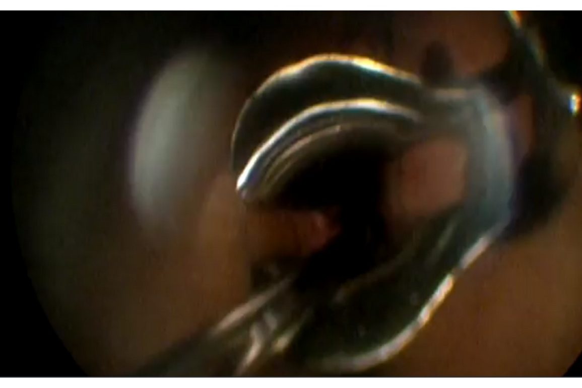
Video Description

This short video outlines the traditional method of hysteroscopic entry into the endometrial cavity.

Presented By

male doctor avatar
Dr. Barry Sanders

Affiliations

University of Calgary

Watch on YouTube

Click here to watch this video on YouTube.

What is Traditional Hysteroscopic Entry into the Endometrial Cavity?

Traditional hysteroscopic entry into the endometrial cavity involves the use of a hysteroscope, a thin, lighted tube, to directly visualize and access the inside of the uterus. The procedure is typically performed as follows:

  1. Preparation: The patient is placed in the dorsal lithotomy position and administered either local, regional, or general anesthesia, depending on the case.
  2. Speculum Insertion: A speculum is inserted into the vagina to hold it open and provide access to the cervix.
  3. Cervical Dilation: If necessary, the cervix is gently dilated to allow the passage of the hysteroscope.
  4. Insertion of the Hysteroscope: The hysteroscope is inserted through the cervix into the uterine cavity. Saline or another distension medium is infused to expand the uterine cavity, allowing for better visualization of the endometrial lining.
  5. Inspection and Procedure: The endometrial cavity is inspected for any abnormalities such as polyps, fibroids, or other intrauterine pathologies. Diagnostic or operative procedures, such as biopsy or removal of abnormal tissue, can be performed under direct visualization.

Traditional hysteroscopic entry is a minimally invasive procedure used for both diagnostic and therapeutic purposes, offering clear visualization of the uterine cavity and targeted treatment of various uterine conditions.

What are the Risks of Traditional Hysteroscopic Entry into the Endometrial Cavity?

The risks of traditional hysteroscopic entry into the endometrial cavity include several potential complications related to the procedure itself and the patient’s overall health. Key risks include:

  • Uterine Perforation: The hysteroscope or surgical instruments can accidentally create a hole in the uterine wall, potentially causing injury to surrounding organs such as the bladder or bowel, which may require additional surgical repair.
  • Infection: There is a risk of infection in the uterine cavity or surrounding areas, which may necessitate antibiotic treatment.
  • Bleeding: Excessive bleeding can occur during or after the procedure, sometimes requiring further medical intervention or, in rare cases, a blood transfusion.
  • Fluid Overload: The use of distension media to expand the uterine cavity can sometimes lead to fluid overload or electrolyte imbalances, particularly if large volumes are absorbed into the bloodstream.
  • Adhesion Formation: Post-surgical adhesions (scar tissue) can form within the uterine cavity, potentially leading to complications such as chronic pain, menstrual irregularities, or infertility (a condition known as Asherman’s syndrome).
  • Reaction to Anesthesia: Complications from anesthesia, such as allergic reactions, respiratory issues, or cardiovascular problems, can arise, especially in patients with underlying health conditions.
  • Incomplete Procedure: There is a possibility that not all abnormal tissue is successfully removed or that the intended diagnostic or therapeutic objectives are not fully achieved, necessitating additional procedures.

Patients undergoing traditional hysteroscopic entry should be fully informed of these risks and receive comprehensive preoperative and postoperative care to minimize complications and support successful outcomes.

Video Transcript: Traditional Hysteroscopic Entry into the Endometrial Cavity

This video demonstrates traditional hysteroscopic entry into the endometrial cavity. An open-sided speculum has already been inserted into the vagina to expose the exocervix. A single-tooth tenaculum is placed on the anterior lip of the cervix to stabilise the cervix. The hysteroscope is placed into the exocervix and the distension media should now be infusing. Using gentle, small movements of the hysteroscope, the channel of the endocervical canal should be brought into view.

Then the hysteroscope should be slowly advanced, while following the canal visually until the endometrial cavity is entered. Once entered, the cavity will fill with distension media and the field should clear with circulation of the distension media. The anatomical landmarks can then be seen clearly. Use the angle of the lens to get a panoramic view of the entire endometrial cavity.