
Dr. Lacey Brennan
Dr. M Jonathon Solnik
Dr. Caroline Leps
Affiliations
Mountain Sinai & Woman College Hospital
University of Toronto
Watch on YouTube
Click here to watch this video on YouTube.
What is Uterine Applications of the Laparoscopic Ultrasound?
What are the Risks of Uterine Applications of the Laparoscopic Ultrasound?
Video Transcript: Uterine Applications of the Laparoscopic Ultrasound
Uterine applications of the laparoscopic ultrasound. We have no relevant disclosures. The objectives for this video are to review the aetiology and management of uterine fibroids, demonstrate the use and setup of a laparoscopic ultrasound for myomectomy, and discuss other uterine applications of a laparoscopic ultrasound, including nontubal ectopic pregnancies.
Leiomyomas, more commonly referred to as fibroids, are benign monoclonal tumours arising from uterine smooth muscle. Prevalent in 70 to 80% of women by the age of 50, common symptoms include menstrual abnormalities, bulk symptoms, pain, and fertility concerns.
Management options for fibroids includes both expectant and medical management, minimally invasive nonsurgical interventions, and finally, traditional surgery, the latter of which will be the focus of this video.
The surgical approach to uterine fibroids is dependent on the goals of treatment, individual patient considerations, clinical factors, including size and location of fibroids, and surgeon comfort with the uterine size, and the number, size, and location of fibroids. Approaches may include hysteroscopic, laparoscopic, or open myomectomies, as well as hysterectomy.
When compared to laparotomy, the laparoscopic myomectomy confers several advantages for appropriately selected patients, including shorter recovery times, improved haemostasis, less post-operative pain, and fewer overall complications.
The ability of the surgeon to accurately detect and remove fibroids in a laparoscopic myomectomy is dependent on factors like preoperative imaging, intraoperative visualisation, laparoscopic ergonomy, and tactile feedback. While robotic-assisted laparoscopic myomectomy may allow for removal of larger fibroids and improved angles for dissection, the tactile feedback is significantly reduced.
Intraoperative laparoscopic ultrasound has been shown to address some of these limitations by providing real time mapping of fibroids, helping identify additional fibroids that may otherwise have been missed. In a prospective study of 42 patients, using a laparoscopic ultrasound permitted detection of a median of two additional fibroids per patient, most commonly type two and three. This is clinically relevant as type two and three fibroids often impact fertility and contribute to heavy menstrual bleeding.
To begin, a 12 mm port is inserted into the patient’s abdomen to facilitate insertion and manipulation of the ultrasound. The pro is then fed into the operative field. Real time ultrasound footage can be seen directly on the laparoscopic screen or on a separate screen, depending on individual provider setup.
Our patient was a 38-year-old G2P0 presenting with heavy menses and infertility. She previously underwent laparoscopic myomectomy with some improvement, but her symptoms returned and were unable to be controlled with medical management. As such, the decision was made to proceed with myomectomy.
After removing the easily visualised dominant fibroid, the laparoscopic ultrasound was used to identify any remaining fibroids. First, we scanned in the coronal plane, then along the anterior wall of the uterus and the sagittal plane, followed by the posterior wall in the sagittal plane. As demonstrated, these manoeuvres permitted us to identify one additional fibroid. Having identified the additional fibroid, the site of the first myomectomy was then closed.
The uterus was then lifted out of the pelvis, and the serosa over the second fibroid was incised, allowing us to perform the second myomectomy. We then close the myometrium and check for haemostasis.
While laparoscopic ultrasound has shown to be a promising tool for myomectomies, there are many other uterine and extrauterine applications that have been documented in gynaecologic literature.
In our case, a 31-year-old G3P1 was referred for management of an ectopic pregnancy. Both outside ultrasound and internal MRI demonstrated concern for left interstitial ectopic. After extensive review with a gynaecologic radiologist, the pregnancy was thought to be either within the left interstitium or intramurally located near the fundus. The laparoscopic ultrasound was there for use intraoperatively for surgical planning.
Upon entry, significant bulging myometrium within the left uterine cornua was clearly visualised. The laparoscopic ultrasound was then used to identify the endometrial cavity in its relation to the pregnancy, allowing us to clearly delineate the borders of the gestational sac. We then began performing a wedge resection of the cornua to ensure complete removal of the pregnancy.
As we dissected through the myometrial layers, products of conception became clearly visible. At this point, we were not sure whether the endometrium was being visualised, or whether there was further extension of the products of conception. The laparoscopic ultrasound was then used again to confirm the location of the endometrial cavity and ensure that the gestational sac in our dissection was appropriately lateral to this.
We then completed the cornual resection. As you can see, we were able to skive the endometrial cavity without entering it. A multilayered repair was then used to reapproximate the overlying myometrium.
In conclusion, the laparoscopic ultrasound is an easy-to-use tool that can aid in surgical planning and precision for uterine surgeries. It can be safely used to identify additional fibroids that may otherwise be missed in laparoscopic myomectomies in order to improve fertility outcomes and reduce heavy menstrual bleeding. Laparoscopic ultrasound can also be used to delineate borders of interstitial and other non-tubal ectopic pregnancies, limiting the amount of healthy uterine tissue resected.
Table of Contents
- Procedure Summary
- Authors
- Youtube Video
- What is Approach to a Uterine Applications of the Laparoscopic Ultrasound?
- What are the Risks of Uterine Applications of the Laparoscopic Ultrasound?
- Video Transcript
Video Description
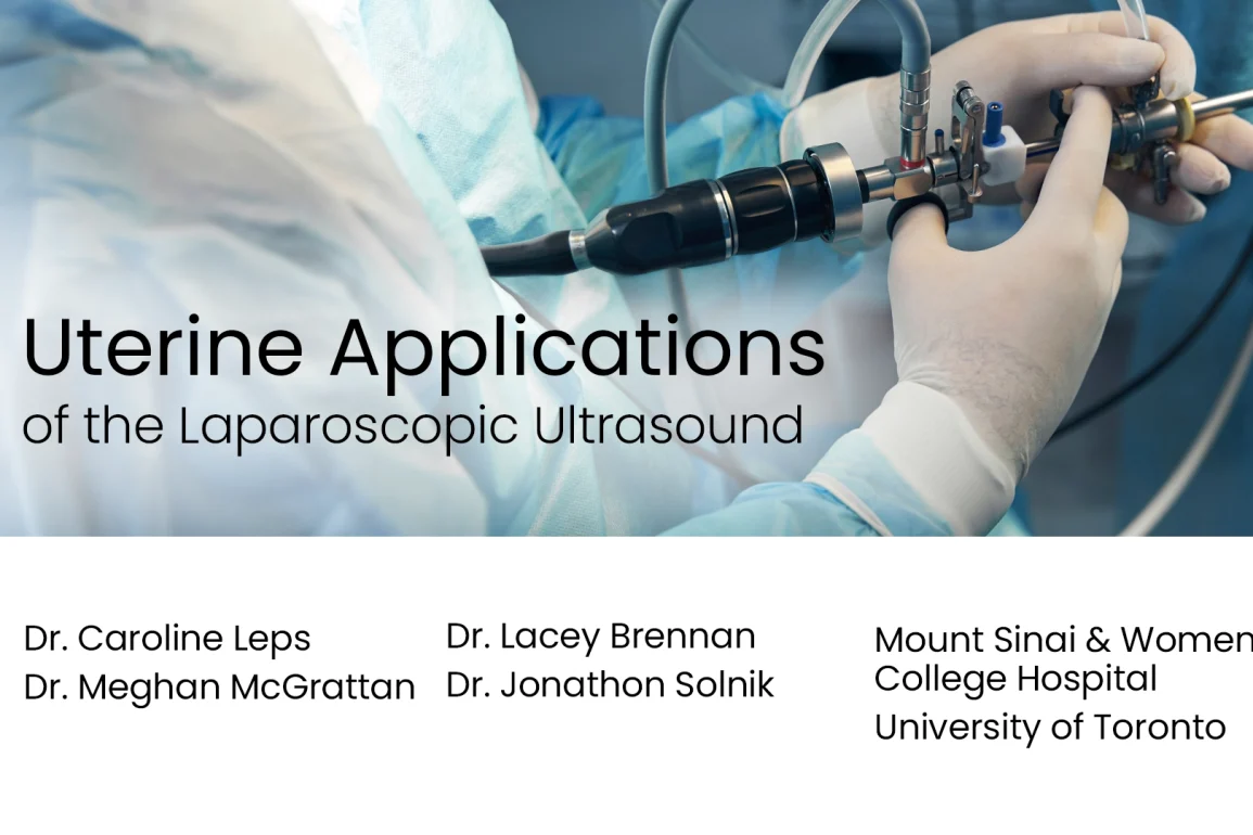
The objective of this video is to demonstrate the use and setup of a laparoscopic ultrasound, show its application to two uterine surgeries, and review accompanying literature. Firstly, we demonstrate the laparoscopic ultrasound’s use during a laparoscopic myomectomy. A recent study has demonstrated that the use of laparoscopic ultrasound permits a median of two additional fibroids to be found per patient, the most common being Grade 2 and 3. These fibroids are of clinical relevance, as they may impact fertility and cause heavy menstrual bleeding. We then demonstrate the use of a laparoscopic ultrasound during the resection of an interstitial pregnancy. This shows how the laparoscopic ultrasound helps permit the minimum amount of healthy uterine tissue to be resected, by clearly delineating the borders of the gestational sac. Other gyneacologic uterine and non-uterine applications are also briefly discussed.
Presented By


Dr. Lacey Brennan
Dr. M Jonathon Solnik
Dr. Caroline Leps
Affiliations
Mountain Sinai & Woman College Hospital
University of Toronto
Watch on YouTube
Click here to watch this video on YouTube.
What is Uterine Applications of the Laparoscopic Ultrasound?
What are the Risks of Uterine Applications of the Laparoscopic Ultrasound?
Video Transcript: Uterine Applications of the Laparoscopic Ultrasound
Uterine applications of the laparoscopic ultrasound. We have no relevant disclosures. The objectives for this video are to review the aetiology and management of uterine fibroids, demonstrate the use and setup of a laparoscopic ultrasound for myomectomy, and discuss other uterine applications of a laparoscopic ultrasound, including nontubal ectopic pregnancies.
Leiomyomas, more commonly referred to as fibroids, are benign monoclonal tumours arising from uterine smooth muscle. Prevalent in 70 to 80% of women by the age of 50, common symptoms include menstrual abnormalities, bulk symptoms, pain, and fertility concerns.
Management options for fibroids includes both expectant and medical management, minimally invasive nonsurgical interventions, and finally, traditional surgery, the latter of which will be the focus of this video.
The surgical approach to uterine fibroids is dependent on the goals of treatment, individual patient considerations, clinical factors, including size and location of fibroids, and surgeon comfort with the uterine size, and the number, size, and location of fibroids. Approaches may include hysteroscopic, laparoscopic, or open myomectomies, as well as hysterectomy.
When compared to laparotomy, the laparoscopic myomectomy confers several advantages for appropriately selected patients, including shorter recovery times, improved haemostasis, less post-operative pain, and fewer overall complications.
The ability of the surgeon to accurately detect and remove fibroids in a laparoscopic myomectomy is dependent on factors like preoperative imaging, intraoperative visualisation, laparoscopic ergonomy, and tactile feedback. While robotic-assisted laparoscopic myomectomy may allow for removal of larger fibroids and improved angles for dissection, the tactile feedback is significantly reduced.
Intraoperative laparoscopic ultrasound has been shown to address some of these limitations by providing real time mapping of fibroids, helping identify additional fibroids that may otherwise have been missed. In a prospective study of 42 patients, using a laparoscopic ultrasound permitted detection of a median of two additional fibroids per patient, most commonly type two and three. This is clinically relevant as type two and three fibroids often impact fertility and contribute to heavy menstrual bleeding.
To begin, a 12 mm port is inserted into the patient’s abdomen to facilitate insertion and manipulation of the ultrasound. The pro is then fed into the operative field. Real time ultrasound footage can be seen directly on the laparoscopic screen or on a separate screen, depending on individual provider setup.
Our patient was a 38-year-old G2P0 presenting with heavy menses and infertility. She previously underwent laparoscopic myomectomy with some improvement, but her symptoms returned and were unable to be controlled with medical management. As such, the decision was made to proceed with myomectomy.
After removing the easily visualised dominant fibroid, the laparoscopic ultrasound was used to identify any remaining fibroids. First, we scanned in the coronal plane, then along the anterior wall of the uterus and the sagittal plane, followed by the posterior wall in the sagittal plane. As demonstrated, these manoeuvres permitted us to identify one additional fibroid. Having identified the additional fibroid, the site of the first myomectomy was then closed.
The uterus was then lifted out of the pelvis, and the serosa over the second fibroid was incised, allowing us to perform the second myomectomy. We then close the myometrium and check for haemostasis.
While laparoscopic ultrasound has shown to be a promising tool for myomectomies, there are many other uterine and extrauterine applications that have been documented in gynaecologic literature.
In our case, a 31-year-old G3P1 was referred for management of an ectopic pregnancy. Both outside ultrasound and internal MRI demonstrated concern for left interstitial ectopic. After extensive review with a gynaecologic radiologist, the pregnancy was thought to be either within the left interstitium or intramurally located near the fundus. The laparoscopic ultrasound was there for use intraoperatively for surgical planning.
Upon entry, significant bulging myometrium within the left uterine cornua was clearly visualised. The laparoscopic ultrasound was then used to identify the endometrial cavity in its relation to the pregnancy, allowing us to clearly delineate the borders of the gestational sac. We then began performing a wedge resection of the cornua to ensure complete removal of the pregnancy.
As we dissected through the myometrial layers, products of conception became clearly visible. At this point, we were not sure whether the endometrium was being visualised, or whether there was further extension of the products of conception. The laparoscopic ultrasound was then used again to confirm the location of the endometrial cavity and ensure that the gestational sac in our dissection was appropriately lateral to this.
We then completed the cornual resection. As you can see, we were able to skive the endometrial cavity without entering it. A multilayered repair was then used to reapproximate the overlying myometrium.
In conclusion, the laparoscopic ultrasound is an easy-to-use tool that can aid in surgical planning and precision for uterine surgeries. It can be safely used to identify additional fibroids that may otherwise be missed in laparoscopic myomectomies in order to improve fertility outcomes and reduce heavy menstrual bleeding. Laparoscopic ultrasound can also be used to delineate borders of interstitial and other non-tubal ectopic pregnancies, limiting the amount of healthy uterine tissue resected.


