Table of Contents
- Procedure Summary
- Authors
- Youtube Video
- What is Laparoscopic cerclage at 15 weeks of gestation after trachelectomy?
- What are the Risks of Laparoscopic cerclage at 15 weeks of gestation after trachelectomy?
- Video Transcript
Video Description
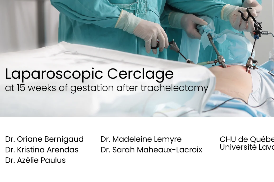
A complex case showing laparoscopic cerclage at 15 weeks gestation following prior trachelectomy, highlighting surgical technique, challenges, and fetal safety.
Presented By
Affiliations
CHU de Québec Université Laval
Watch on YouTube
Click here to watch this video on YouTube.
What is Laparoscopic cerclage at 15 weeks of gestation after trachelectomy?
Laparoscopic cerclage at 15 weeks of gestation after trachelectomy is a fertility-preserving procedure used to prevent pregnancy loss when little or no cervix remains following surgical removal of the cervix for early cervical cancer. A permanent suture is placed around the lower uterus (at the level of the internal os) through a minimally invasive abdominal approach to provide mechanical support to the pregnancy.
- Preoperative Planning: Ultrasound assessment of cervical length and uterine anatomy ensures the safest site for suture placement and guides trocar positioning for the gravid uterus.
- Laparoscopic Entry: Ports are inserted using an open (Hasson) technique and insufflation pressures are kept low to protect the pregnancy.
- Bladder and Broad Ligament Dissection: The vesicouterine peritoneum is incised and the bladder gently dissected downward to expose the uterine isthmus and identify the uterine arteries.
- Suture Placement: A non-absorbable monofilament suture is passed through windows created in the broad ligament on both sides of the uterus, medial to the uterine arteries, encircling the isthmus to provide circumferential support.
- Knot Securing and Peritonealisation: The suture is tied securely using a sliding knot and reinforced with additional knots, then covered with peritoneum to reduce adhesion formation.
This cerclage is typically left in place for the remainder of the pregnancy and for future pregnancies, with delivery by cesarean section.
What are the Risks of Laparoscopic cerclage at 15 weeks of gestation after trachelectomy?
Laparoscopic cerclage during an ongoing pregnancy carries both surgical and obstetric risks.
- Pregnancy Loss or Preterm Labor: Manipulation of the gravid uterus and anesthetic exposure can trigger miscarriage, preterm contractions, or premature rupture of membranes.
- Bleeding: Dissection near the uterine arteries increases the risk of intraoperative hemorrhage requiring transfusion or conversion to open surgery.
- Bladder or Ureteral Injury: The bladder and ureters lie close to the dissection plane and may be damaged during peritoneal or broad ligament dissection.
- Infection: Pelvic or port-site infection can occur, potentially threatening the pregnancy.
- Anesthetic Complications: Laparoscopy during pregnancy requires careful control of insufflation pressures and maternal positioning to avoid fetal compromise.
- Future Delivery Limitations: Because the suture is permanent, all future births must be by cesarean section.
Meticulous surgical technique, multidisciplinary planning, and careful intraoperative monitoring help minimize these risks while providing strong cervical support to maintain the pregnancy.
Video Transcript:
The Minimally Invasive Gynaecologic team at Laval University in Quebec present a technique for abdominal cerclage at 15 weeks of gestation after a trachelectomy. Here are some key tips and tricks for abdominal cerclage during pregnancy. We present a post-trachelectomy case at 15 weeks, and highlight the key surgical steps. Nearly all of women diagnosed with cervical cancer are younger than 45 years, with a significant proportion wishing to preserve their fertility.
Currently, for tumours less than two centimetres, fertility-sparing surgery is considered safe, and is offered as part of standard practice. Moreover, in cases of frequent miscarriages, preterm birth, or failure of vaginal cerclage, the [unclear] recommends that women who have undergone a trachelectomy should receive an abdominal cerclage.
Additionally, the placement of an emergency cerclage may be considered for women whose cervix is dilated less than four centimetres without contractions, and who are under 24 weeks pregnant.
The patient is 37 years old, with a history of trachelectomy. This is her second pregnancy achieved through IVF, following a spontaneous early miscarriage. At 15 weeks of gestation the cervical length measured 17 mm with insufficient cervical tissue to allow for a vaginal cerclage.
Step one, preparation with a gravid uterus. To obtain adequate exposure, the assistant on the patient’s right uses a liver retractor designed to be non-traumatic for the gravid uterus. The operating table tilt is adjusted as needed throughout the procedure. We performed a Hasson open technique to insert the laparoscope, minimising the risk of uterine trauma.
Two 5 mm circles were placed in the iliac [unclear], and one 11 mm trocar was positioned three finger width above the umbilicus. Insufflation pressures do not exceed 15 mm of mercury.
In the context of pregnancy, monopolar current is avoided in favour of advanced bipolar energy. Additionally, the patient received Indocin, and was positioned with [unclear] compression stockings.
Step two, we want to expose the isthmus and the uterine artery. First, the vesicouterine peritoneum was inside, and the anterior leaf of the broad ligament was opened. The liver retractor continued to provide exposure in the least traumatic manner possible. The bladder was then dissected, and gently reflected inferiorly using blunt dissection.
The posterior leaf of the broad ligament was fenestrating using a grasper through the major trocar. [Unclear] incision is [unclear] caudally by blunt dissection. The same was done on the right side. Tilted [unclear] are key to optimise exposure air, and equally important during cerclage placement. Following this dissection steps, the uterine artery was [unclear] along its [unclear] course.
Step three, placement of the cerclage. A non-absorbable monofilament suture made of polypropylene, size one, and 75 cm in length was used. Identify the isthmus. Gentle pressure in the anterior fornix can help determine the correct level for cerclage.
Avoid traditional manipulators. Insert the needle between the uterus and the uterine artery. Some bleeding is common. [Unclear], it usually stops when the cerclage is tied. [Unclear], the previously fenestrating left broad ligament, encircling the uterine fundus.
Notably there were no adhesions in the posterior compartment. The suture was then passed through the window, in the right broad ligament. Finally, the needle was passed again, medial to the uterine artery. One or two sutures can be placed at [unclear] isthmus.
Step four, the roder knot and peritonealisation. The roder knot eases the application of the right pressure. It allows sufficient tension without tissue strangulation, or ischaemia. It is then secured with four additional intracorporeal knots. Then we confirm that the suture is placed at the correct level. Peritonealisation of the cerclage suture was performed using a band suture.
The vesicouterine peritoneum is carefully closed to seal the fenestration of the broad ligament. Since [unclear] are more prominent during pregnancy, it’s important to use a haemostatic agent if bleeding occurs. At 36 weeks the patient underwent a C-section following premature [unclear], and delivered a healthy newborn. The cerclage was left in place for the following pregnancy.
In conclusion, due to the limitations imposed by the gravid uterus, carefully planning is required for optimal exposure. In step one, we begin by placing the trocars higher, using a Hasson open technique. Insufflation is limited to 15 mm of mercury, and monopolar energy is avoided.
A hepatic retractor ensures gentle and safe exposure. Step two, next we dissect the vesicouterine peritoneum, and both leaves of the broad ligament, to identify the uterine arteries.
Moving on to step three, using a vaginal manipulator, a monofilament suture is [unclear] to the uterine arteries. The cerclage tension helps stop any bleeding. Finally, in step four, with the cerclage in place, a roder knot [unclear] suture, followed by four intracorporeal knots.
A peritonealisation of the cerclage is performed. Let note that according to the literature it may be more optimal to perform this technique outside of pregnancy for better pelvic exposure, as well as to avoid surgery under general anaesthesia during pregnancy. I would like to thank the [unclear] team for the cooperation. Thank you for watching.


