Table of Contents
Summary
- Indications for appendectomy in gynecology include epithelial ovarian cancer, mucinous ovarian neoplasms, pseudomyxoma peritonei, anticipated abdominal or pelvic radiation, and chronic pelvic pain.
- The appendix is located in the right lower quadrant, and its blood supply is mainly from the appendicular artery.
- The surgical procedure for appendectomy involves mobilizing the appendix, isolating and dividing the appendiceal artery, and placing endoloops at the base of the appendix to prevent complications.
- Complications of appendectomy may include bleeding, intra-abdominal abscess, wound infection, and unrecognised enteric injury.
- Patients should be advised to seek immediate attention if they experience significant pain, fevers, vomiting, or other concerns after the procedure.
Presented By
Affiliations
See Also
Appendectomy Explanation
- An appendectomy is a surgical procedure to remove the appendix, a small, tube-like organ attached to the large intestine.
- It’s often performed as emergency surgery to treat appendicitis, an inflammation of the appendix typically caused by a blockage leading to infection.
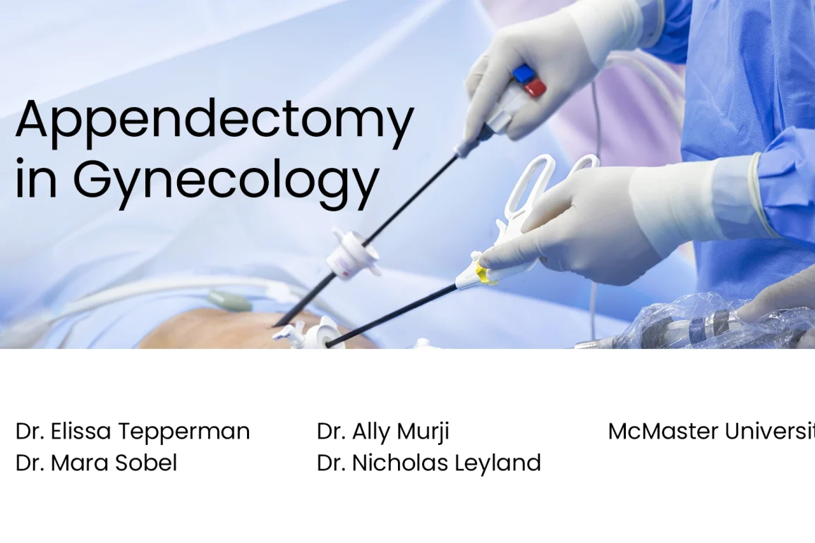
- The surgery can be done as an open surgery (single long incision) or laparoscopic surgery (multiple small incisions and the use of a camera for guidance).
- Post-surgery recovery typically occurs within a few weeks, but this depends on the individual’s health and the severity of the appendicitis.
- Possible complications include infection, bleeding, hernia at the incision site, blocked bowels, or injury to nearby organs.
- The exact function of the appendix is unknown, but people can live without it without any apparent consequences.
Watch on YouTube
Click here to watch this video on YouTube.
Video Transcript
Appendectomy is one of the most common operations performed by general surgeons. From a gynaecologic perspective, there are important additional considerations for removal of the appendix. And this straightforward operation may be performed in a safe, timely and cost-effective manner in the hands of a properly trained gynaecologic surgeon. This video will review the gynaecologic indications for appendectomy, the relevant anatomy, the surgical procedure and complications of appendectomy that the gynaecologist should be aware of.
Gynaecologic Indications for Appendectomy
Appendectomy is indicated in cases of epithelial ovarian cancer, mucinous ovarian neoplasms and pseudomyxoma peritonei. Appendectomy may be considered in cases of anticipated abdominal or pelvic radiation, or where extensive pelvic or abdominal surgery may incur severe post operative adhesions. Finally, removal of the normal or abnormal appearing appendix may be considered in patients with chronic pelvic pain and or right lower quadrant pain as appendiceal pathology is common in this patient population.
Appendectomy in Cases of Endometriosis
Similarly, appendectomy may be considered in cases of endometriosis, irrespective of a normal or abnormal appearing appendix due to the high association of pathology. Specifically, appendectomy at the time of ovarian endometrioma resection should be considered as this finding is associated with deeper intestinal disease. This is an example of a dysmorphic appendix seen at laparoscopy in a woman with endometriosis.
Here we see the characteristic hockey stick appearance with the appendix having a curved distal end. No preoperative symptoms or clinical findings predict involvement of the appendix in women with endometriosis. The rationale for performing incidental appendectomy in these women includes the elimination of future appendicitis, or diagnostic consideration of appendicitis and the removal of undiagnosed incidental pathology involving the appendix.
The Anatomy of Appendix
The appendix is located in the right lower quadrant. It is a blind ended tube connected to the cecum, its embryologic origin. The human appendix averages 10 cm in length, but can range from 2 to 20 cm. The diameter of the appendix is usually between 7 and 8 mm. While the base of the appendix is at a fairly constant location 2.5 cm below the ileocecal valve on the posteromedial wall of the cecum, the location of the tip can vary in position, including retrocaecal, pelvic or extraperitoneal.
Appendix Blood Supply
Blood supply to the appendix is mainly from the appendicular artery which is a branch of the ileocolic artery. This artery courses through the mesoappendix posterior to the terminal ileum, and accessory appendicular artery can branch from the posterior cecal artery. Unrecognised damage to this vessel may lead to significant intraoperative haemorrhage.
Appendectomy Surgical Procedure
The patient is placed in steep Trendelenburg position until the small bowel remains above the pelvic brim. This maximises exposure of the pelvis and large bowel. Here the cecum and appendix are visualised. Although the appendix may be directly grasped within atraumatic laparoscopic instrument, we prefer to manipulate the appendix via the mesoappendix to minimise bowel injury. In some cases, where parts or all of the appendix lies retroperitoneally releasing the peritoneum between the cecum and lateral sidewall will optimise visualisation.
Steps for Appendectomy
If adhesions are present, these should be liased [?] as complete mobilisation of the appendix is essential. Note that here we are using an umbilical video laparoscope and three ancillary ports, although any port configuration which allows triangulation with the appendix is appropriate. Once the entire length of the appendix is mobilised, the appendiceal artery within the mesoappendix is isolated near the base of the appendix. Here we use the harmonic scalpel to carefully dissect the fatty mesentery of the appendix.
Complications in Appendectomy
Next, the appendiceal artery is sealed, then divided again using a harmonic scalpel with excellent haemostasis. Alternatively, vascular clips may be used instead for this step. Any remaining mesentery is dissected towards the base until the appendix is completely free and devascularised. Two endoloops are placed at the base of the appendix, flushed with the cecum, to prevent residual tissue and the associated complications such as stump appendicitis. These endoloops will remain in situ.
Next, we gently melt the appendix content towards the tip before securing a third endoloop just distilled to the previous endoloops in order to seal the appendix prior to removal. The appendix is then cut between the endoloops and placed within a specimen bag which is removed through the umbilical port and sent to pathology. Alternatively, an automatic tissue stapler can be used for this step. Irrigation and inspection of the appendiceal pedicle under low pressure reveals excellent haemostasis.
Aftercare
The most common intraoperative complication is bleeding. Injury to an accessory appendicular artery can lead to intraoperative haemorrhage and therefore careful inspection for haemostasis under low intra-abdominal pressures is advised prior to closure. Intra-abdominal abscess is rare since our patients typically do not have an acutely inflamed appendix at the time of surgery. Wound infection is a more common complication and is minimised by administering intraoperative antibiotics. Unrecognised enteric injury is serious and potentially life threatening.
Patients should be clearly counselled to seek immediate attention should they develop significant pain, fevers, vomiting or other concerns. There are several important gynaecologic indications for appendectomy. A knowledge of the relevant anatomy and management of complications are important for the gynaecologist to safely perform this procedure. Alternatively, a consultant general surgeon may be called upon to perform the procedure according to the gynaecologist comfort and preference.


